Hello friends, in this article, we will learn what the plasma membrane is and also learn about it, such as its definition, structure, chemical composition, functions, etc. So let’s start without wasting time.
What is a Cell membrane or plasma membrane?
Definition –
The plasma membrane is the outer covering of the cytoplasm of the cell. It acts as a selective filter and controls the entrance and exit of molecules and ions, thus helping maintain the difference in the cytoplasm’s ionic concentration and surroundings.
Ultrastructure of the plasma membrane?
The plasma membrane is about 75 angstroms thick, But its thickness varies in different types of cells and ranges from 75 to 105 Angstroms or up to 215 angstroms. Under the electron microscope, 3 distinct layers could be differentiated in the plasma membrane.
- How to have a dense layer of protein: about 20 to 25 Angstroms.
- Middle bimolecular layer Of Lipids: about 30 – 35 Angstroms.
- The inner dense layer of protein: about 20-25 Angstroms.
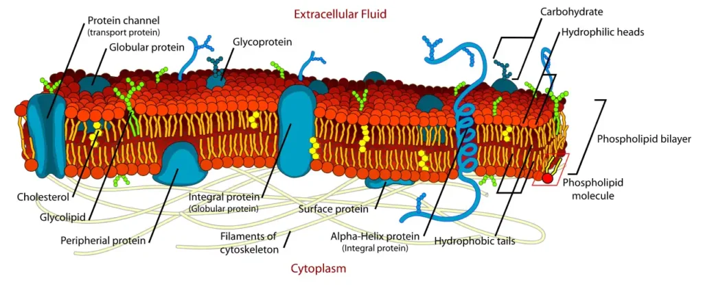
The plasma membrane consists of a bimolecular layer of lipids sandwiched between a Monomolecular protein layer. Robertson has described such a membrane as a unit membrane.

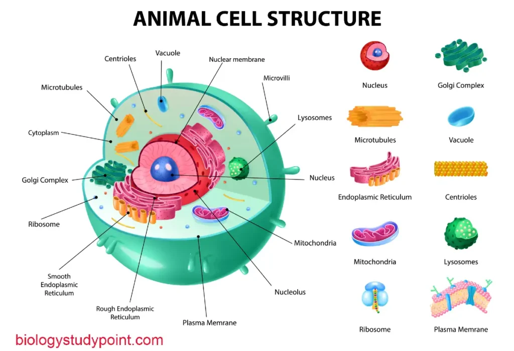
He found that various cell structures like endoplasmic reticulum, Golgi complex, nuclear membrane, mitochondria, plastids, and lysosomes are surrounded by a unit membrane.
The Chemical composition of the cell membrane or plasma membrane
The plasma membrane of animal cells consists of proteins, lipids, and a few carbohydrates.
Proteins –
The protein is the main bulk of the plasma membrane. The protein is separated from the plasma membrane of R.B.C.They are collectively known as tektins. These are of two different types.
Peripheral or extrinsic proteins –
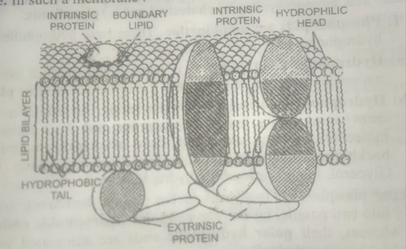
These form the outer and inner layers of the middle Lipid layer of the plasma membrane. These are loosely connected and timely separated even by milled treatment. These are soluble in aqua solution.
Spectrin in erythrocytes, cytochrome C in mitochondria, and acetylcholinesterase in electroplate membranes are peripheral proteins.
Integral or intrinsic proteins –
These proteins Penetrate the lipid layer partially or wholly. Their polar heads protrude from the surface of the membrane, while the nonpolar regions are embedded in the interior of the membrane.
These are insoluble in water. Some detergents or organic solvents are required to separate them from the membrane.
These are attached to the oligosaccharides (glycoproteins) or phospholipids (proteolipids or lipoproteins).
Lipids
The plasma membrane contains about 20 – 40% of lipids. The lipid component is of three types –
- Phospholipids like lecithin, serine, cephalin, and sphingomyelin;
- Cholesterol and
- Glycolipids.
- Phospholipids are amphipathic. These have hydrophilic heads and hydrophobic tails. The hydrophobic head end is a water-loving part of phospholipid molecules called the polar end. It is formed of choline phosphate.
- Hydrophobic tail end – It is a water-hating part that moves away from water and is known as the nonpolar end. It is formed of two molecules of fatty acids. Both these molecules are joined to the backbone of glycerol through their carboxyl group (-COOH). Glycerol, in turn, is joined to the hydrophilic head,
These phospholipid molecules in the plasma membrane are arranged radially into two parallel layers. The nonpolar hydrophobic ends face Each Other, whereas their polar hydrophilic ends are associated with protein molecules or carbohydrates.
2. Cholesterol
Cholesterol is found in Eukaryotic cells’ plasma membrane and other intracellular membranes. It is usually present in the tails of phospholipid molecules. It provides rigidity to tables and mechanical stability to the lipid bilayer.
Glycolipids are found only in the outer lipid layer. These carry carbohydrate chains.
What are the functions of lipids?
- The lipid layer forms the structural framework of the plasma membrane.
- It forms a permeability barrier to the ions and polar molecules entering the cell.
3. Carbohydrates
The plasma membrane contains 2 to 10% carbohydrates, which occur in two different combinations: hexose, hexosamine, fucose, and sialic acid.
- Attached to lipids – glycolipids;
- Attached to proteins – glycoproteins.
Fluid mosaic model of cell membrane
The fluid mosaic model was proposed by Singer and Nicholson (1972).
According to this concept, all biological membranes have quasifluid structures in such a membrane.
- The lipids and integral proteins in the plasma membrane are amphipathic, containing both hydrophobic and hydrophilic groups. They do not have polar groups inside their hydrophobic bilayer; the hydrophilic groups are directed towards the water phase.
- Lipids and integral proteins are arranged in a mosaic fashion.
- The lipid-protein and Oligosaccharides are held together in the plasma membrane by noncovalent interaction (Gitler 1972).
- The membrane’s fluidity is associated with the fluidity of lipids, which in turn depends on the degree of saturation of hydrocarbon Chains of lipids. Unsaturated fatty acids having double or triple bonds have a lower melting point than saturated ones. Most biological membranes have sufficient and saturated lipids so that the lipids by layers are quasi-fluid at Body temperature. Cells living at low temperatures have a higher proportion of unsaturated fatty acid than cells living at higher temperatures.
- The liquid and protein molecules can perform lateral movement within the lipid bilayer. The lipid molecules may show (i) an intramolecular moment (i.e., motion with lipid molecules), or (ii) they may rotate about their axis. (iii) rarely do these exhibit ‘flip-flop’ moments, which include transfer from one side of the bilayer to the other.
- The integral protein exhibits three different types of movement inside the fluid layer: (i) Lateral diffusion, (ii) rotational diffusion around an axis perpendicular to the surface of the membrane, and (iii) rotational diffusion around an axis parallel to the surface of the membrane.
- Because of such an arrangement of molecules, the active sites of enzymes and antigenic glycoproteins lie on the outer surface of membranes.
- Because of the fluid structure, Protein macromolecules can diffuse through the plasma membrane.
What are Pinocytosis and Phagocytosis?
Pinocytosis or Endocytosis (Cell Drinking)
Pinocytosis in the uptake of fluid. It was first observed by Lewis (1931) and confirmed by Mast and Doyle (1934). Protein amino acids and certain ions induce pinocytosis and occur in white blood corpuscles, Kidney cells, cells of the intestinal mucosa, and hepatic cells.
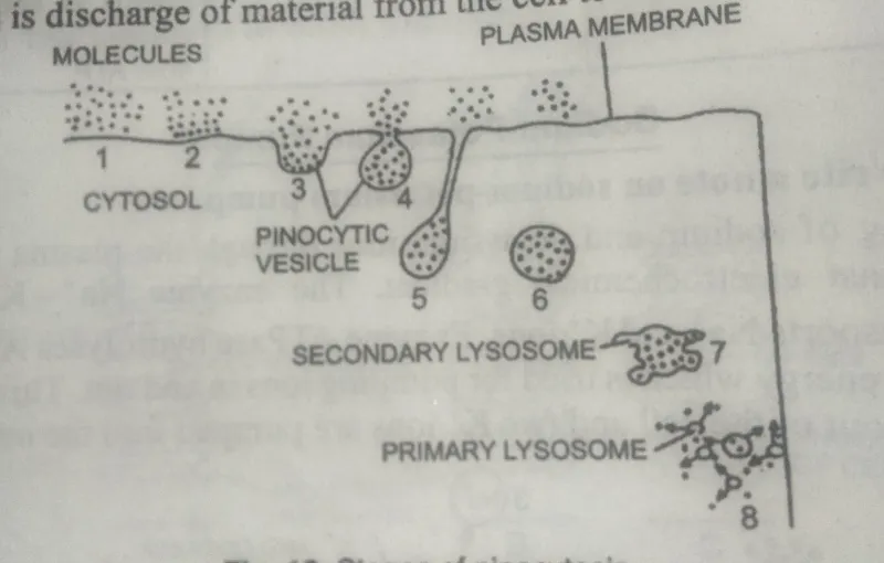
Proteins are amino acid molecules in the medium outside the cell that bind to the specific reporter in the plasma membrane. At this point, the membrane invaginates and produces a cup-like depression in the pinocytic vesicle.
The pinocytic vesicle is pinched off the plasma membrane and into the cytoplasm, which is called a pinosome. It migrates deeper inside the cell, fuses with the primary lysosome, and forms a secondary lysosome. The lysosomal enzymes digest the substance, releasing the digested material into the cell cytoplasm.
Phagocytosis (Ingestion of solid material)
Phagocytosis is engulfing large particles of solid matter through a plasma membrane. It is found in protozoa granular leukocytes, macrophages of the reticuloendothelial System, and the endothelial cells lining the capillary sinusoids of the liver, adrenal gland, and hypophysis.
Phagocytosis is a form of feeding in protozoa, but it is a means of Defense against foreign organisms in metazoa.
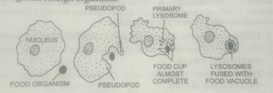
During phagocytosis, the particle is an organism that adheres to the plasma membrane (adsorption) and induces the cell to extend pseudopodia. These pseudopodia Surround the particle and form a food cup.
Finally, the pseudopodia fuse insert links with the particle to form a phagocytic vacuole or phagosome. The phagosome fuses with the primary lysosome to create a secondary lysosome, where the material is digested or degraded.
What is Exocytosis (Cell Vomiting)?
Secretory cells release the secretory contents to the exterior. Releasing substances from the cell to the outside is called exocytosis or cell vomiting.
The undigested material from the phagocytic vacuoles or pinocytic vesicles of the cell is also expelled by exocytosis. The process may involve any of the following routes:
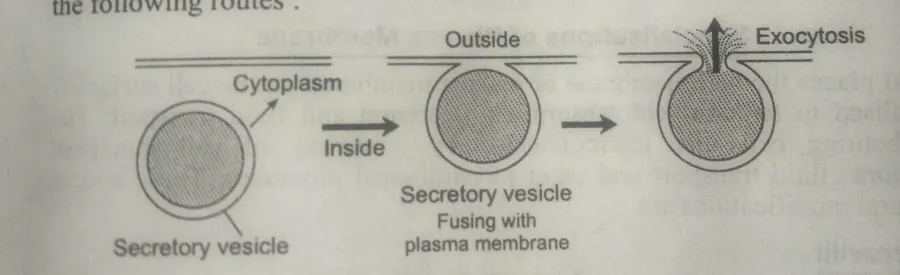
- Secreted molecules may stick to the inner surface of the plasma membrane or Cell Membrane and be expelled from the cell.
- The secretory product is collected in the channels of the Endoplasmic reticulum. It passes through the Golgi complex, gets concentrated, and is packed into secretory vesicles separate From the Golgi complex, moves toward the plasma membrane and fuses with it, and the contents of the vesicle are released outside.
What are the functions of the cell membrane or plasma membrane?
- Regulation of the passage of materials or selective transport of substances
The plasma membrane is selectively permeable. It facilitates the entrance of needed nutrients into the cells and permits the exit of nitrogenous wastes.
The exit of substances from the cell. Thus, the cell membrane regulates the passage of materials into And out of the cell. The transport of materials into and out of the cell occurs through.
It includes the transport of substances by diffusion osmosis and facilitated diffusion.
(ii) Active transport
Active transport in Tamil movement of molecules against a concentration gradient,i.e., the molecule or ions move from the region of low concentration towards the region of high concentration.
The moment of molecules can be compared with the uphill movement of water. Therefore, energy is required during active transport. ATP provides this energy.
(iii) Endocytosis
It includes pinocytosis and phagocytosis, in which solid materials are taken.
(iv) Exocytosis
It expels the waste or cell secretion.
2. Maintenance of differential Distribution of Ions
The plasma membrane controls and maintains the different distribution of Ions inside and outside the cell.
For example, potassium ions are concentrated inside a living cell, whereas Sodium and chloride ions are distributed outside. Regardless of variation in the concentration of iron, the same pattern of a symmetrical distribution across the plasma membrane always occurs.
This unequal distribution of ions leads to a potential difference. This electric energy in all living cells is known as the membrane potential.
This distribution of ions between the inside and outside of the living cell is in a state of low entropy. Therefore, a cell needs to spend a large amount of energy to maintain this differential distribution.
If a cell is poisonous in such a way that it cannot produce energy molecules, the different distribution of Ions is destroyed. The loss of metabolism destroys membrane potential. Interference in membrane potential results in the loss of energy metabolism.
3. Response to the environment
The plants respond to changes in their environment with the help of receptor proteins. These receive chemical messages from the other cells and thus help in cell recognition. The receptor proteins respond to hormone growth factors and neurotransmitters.
4. Contact with neighboring cells
The plasma membrane maintains structural and chemical relationships with the neighboring cells. The glycoproteins of plasma membranes help recognize, maintain communication, and exchange material.
5. Cell Recognition
Cells of the body’s defense system recognize the self and the self and recognize foreign intruders like bacteria, etc., which help in self-protection. The glycocalyx (glycolipids) on the surface of the plasma membrane provide molecular identity to the cells.
6. Communication between neighboring cells
Animals communicate with the cells through gap Junctions. Salts, amino acids, sugar, vitamins, hormones, etc., pass through Gap junctions. Hormones act as chemical signals between distant cells.
7. Cellular Adhesion
All cells of the body remain in close association with other cells. There are several types of cell contacts and adhesion.
8. Protection
A plasma membrane protects the cell and may be involved in a moment of cell secretion and transmission impulses.
9. Excretion
Plasma membranes permit the exit of nitrogenous waste and other waste by diffusion or exocytosis.
Friends, if you like the information, share it as much as possible.
Thank you