Hello, guys. In this article, we are going to study Centrioles. What are centrioles? What are their structure, functions, Composition, etc.? Today, we will learn the answers to many such questions, so let’s start.
What are centrioles?
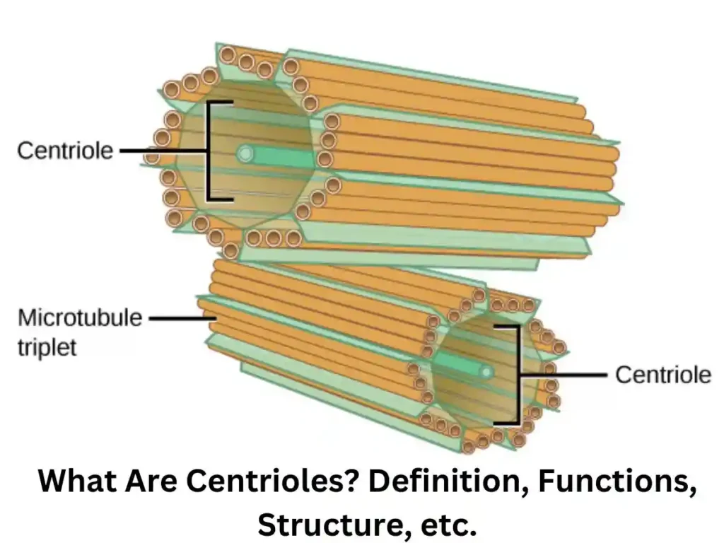
Two spindle-shaped or cylindrical structures are found near the nucleus of the cell. These are called Centrioles, and the two centrioles together are called Centrosomes. They are situated at right angles to each other. A centrosphere surrounds the centrosomes. The hyaline cytoplasm surrounding the Centrosome is called centroplasm.
Centrioles are primarily found in bryophytes, algae, ferns, and all animal cells. They are not found in gymnosperms, red algae, angiosperms, or protozoa without flagella or cilia.
It helps in cell division; let us know how they do it. When cell division occurs, cells form fibers, which are called spindle fibers. Now, it comes to the question that plant cells also do cell division, and the centromere is not found in them. So, how do they do cell division?
Who discovered the Centriole?
The centroid or Centrosome was discovered by two scientists, Bendem and Boveri. Centrioles are membraneless structures.
Centriole is not found in which cells?
Centrioles are not found in nerve cells, mature RBCs, ovum, or plant cells.
Plant cells have a polar cap structure, which forms fibers that help in cell division.
Centrioles Structure
The Centriole’s structure is the same in all cells. Its size ranges from 150 to 250 nm (1500–2500 Å) in diameter and 3000–20000 Å in length.
A centriole is a membrane-less structure and is made up of microtubules. These microtubules are located at equal distances around the center as a circle.
If we talk about its arrangement, it is 9+0. Now, see what 9+0 means. 9+0 means nine micro tubes will be found at the periphery, and no micro tubes will be found in the center. What will we call this 9 + 0 arrangement? When it comes to whether the nine micro tubes on the periphery are doublets or triplets, then the microtubes present here are triplets.
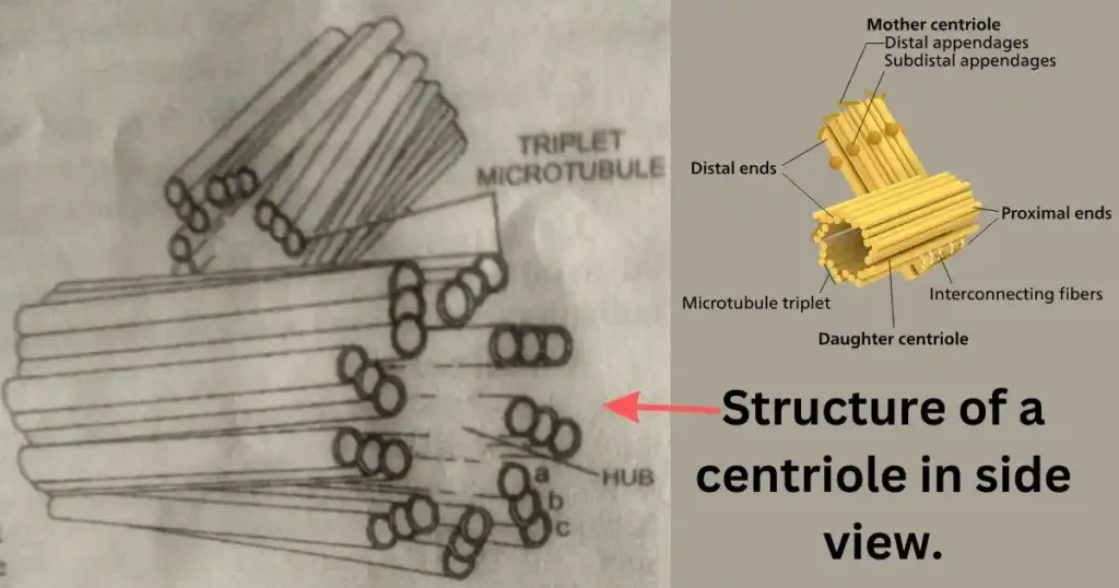
Triplets means the microscopic tubes are in groups of three. The structure of the centrioles formed this way is similar to the cartwheel structure.
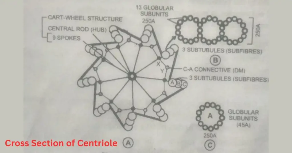
Now, the question is whether these three are interconnected. Yes, all three are connected, and the connecting structure is called a bridge. What happens when there is no microscopic tube in the middle of the Centriole? The center of the centrioles is filled with protein called the hub. The microtube inside this hub is connected to the radial rod.
1. Microtubules –
The triple microtubules present in the Centriole are all the same. Each triplet comprises three subfibres named A, B, and C. The diameter of each subfibre is approximately 250 Å. Subfibre A is tubular; its wall shall consist of 13 globular subunits.
Subfibre B and Subfibre C are C-shaped because B shares three subunits with Subfibre A, and Subfibre C shares three with Subfibre B. The diameter of each subunit is approximately 40–45 Å.
2. Linker’s –
A thick substance connects Subfibre A of each triplet to Subfibre C of the neighboring triplet. But sometimes, this connection can be between A–A or C–C subunits.
Associated Structures (Pericentriolar structures)
Satellites –
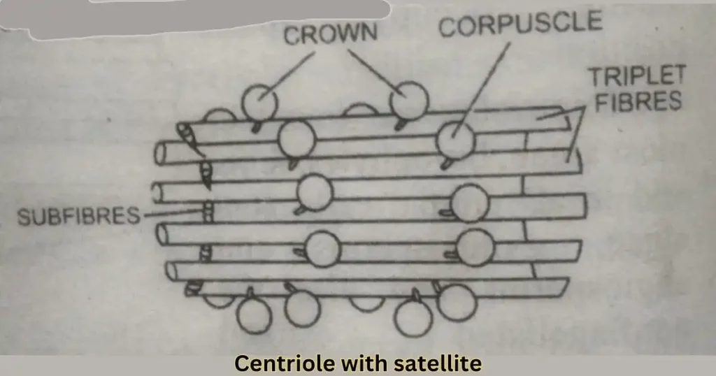
Two crowns surround each Centriole, each consisting of nine amorphous plates or spheres. Small bridges of dense material connect these round structures to the centroid. These structures are called corpuscles, masses, or pericentriolar satellites. The shape, size, and position of satellites change during spermatogenesis.
Biochemical Composition –
Centrioles comprise structural proteins, lipids, tubulin, ATPase, and RNA.
What is the function of Centrioles?
- Centrioles play an essential role in cell division by forming spindles.
- These create the basal bodies of cilia and flagella.
- The distal Centriole in sperm forms the axial filament of the flagellate of sperm.
Centrioles vs Centrosome
The Centrosome, also called the Microcentrum, is the clear zone of cytoplasm surrounding the Centriole. A centriole, a cell center, is a cylindrical body of 9 microtubules or fibers arranged equidistantly in a circle around an imaginary axis. These help spindle formation during cell division and form the basal bodies of cilia and flagella.
Centrosome vs Centromere
The centromere is the region of the chromosome where sister chromatids remain attached. Through the centromere, the chromosome attaches to the equator of the spindle.
Conclusion –
Friends, if you like the centriole information, please share it as much as possible and comment.
Thank you so much