Hello friends, do you want to know everything about epithelial tissue? If yes, then this article is for you only. It will provide complete step-by-step information about Epithelial tissue.
Before reading about epithelial tissue, let’s define tissue. So, without wasting any time, let’s start reading.
What is a tissue?
A tissue is a group of cells with the same function and origin but similar or dissimilar based on structure.
How many types of tissues?
There are four types of tissues in animals.
- Epithelial tissue
- Connective tissue
- Muscular tissue
- Nervous tissue
In this article, we will study only epithelial tissue in detail; the rest will be studied in another article.
Some important questions related to tissue –
Q.1 What is the study of tissue called?
The study of tissue is called Histology.
Q.2 Who gave the word Histology?
The term Histology was given by the scientist Mayer.
Q.3 Who is called the father of Histology?
Marcello Malpighi is called the father of Histology.
Q.4 Who is the father of microscopic anatomy?
Marcello Malpighi is called the father of microscopic anatomy.
Q.5 Who gave the word tissue?
Bichat used the word “tissue” to refer to animals, and Nehemiah Grew used it to refer to plants.
How are the four types of tissues found in animals formed or originated?
All these four tissues are formed from germ layers.
- Epithelial tissue can be formed from any of the three layers of the Ectoderm, the Mesoderm, and the Endoderm.
- Connective tissue is formed only from the Mesoderm layer.
- The formation of muscle tissue also occurs only at the Mesoderm layer.
- Neural tissue is formed only from the ectoderm layer.
What is epithelial tissue?
Epithelial tissue can originate from any of the three layers.
Also, read –
- What is the animal kingdom? Complete notes.
- What is the plant kingdom? Full Information.
- What is biology’s definition and branches?
- What is a cell wall? Complete notes
What is the primary function of epithelial tissue?
Epithelial tissue forms the covering and lining.
Example of covering: Our skin is made up of epithelial tissue. Our skin surrounds the body from the outside, making a cover that works as a protective layer.
Similarly, the lining of the internal cavity (space) inside the body also comprises epithelial tissue. An example of a lining is our alimentary canal. You must have known about it; if you do not, there is no point in taking tension; I am telling you about it.
Our alimentary canal extends from the mouth to the anus. This alimentary canal is divided into different parts. Like – as the mouth, palate (hard palate, soft palate), pharynx, esophagus, stomach, small intestine, large intestine, rectum, and anus.
These are all part of the alimentary canal. This alimentary canal is hollow from the inside; that is, there is a space inside, so you know that the lining of this space is made up of epithelial tissue.
Epithelial tissue can be formed from all three germ levels; we have already read that the part from the mouth to the hard palate is made of Ectoderm and the part from the soft palate to the rectum is made of Endoderm, and from the anal canal to anus Part is made of Ectoderm.
Now, you must ask yourself what makes Mesoderm, the layer of our blood vessels.
Characteristics of epithelial tissue
Epithelial tissue is formed first in our body.
See why this tissue is named Epithelial. Epithelial tissue, of which Epi means upon and upon means “over someone.” Similarly, Thelial means Growth, and Growth means ” increase.”
If these two are read together, it means rising above someone. This means that this tissue grows on someone, and now it comes to the point on whom this epithelial tissue grows. This epithelial tissue grows over the connective tissue; hence, it is called epithelial tissue.
However, connective tissue and epithelial tissue are not adjacent to each other. A layer: A layer called the basement membrane separates these two tissues.
Both tissues make this base layer. How to make it: see the epithelial tissue that secretes a chemical called Glycoprotein on its bottom side, and this chemical forms a layer called Basal lamina.
Similarly, connective tissue secretes a chemical called Mucopolysaccharide on its upper side, forming a Fibrous lamina layer. This layer also contains collagen fibers. These two levels are called the basement membrane at the non-cellular layer.
Epithelial tissue does not have intercellular spaces. This means there is no space between their cells, or their cells are very close to each other.
This tissue is on top of a thin acellular membrane called the basement membrane.
Classification of epithelial tissue –
There are three types of epithelial tissue.
- Simple Epithelium Tissue
- Compound Epithelium Tissue
- Specialized Epithelium Tissue
What is simple epithelial tissue?
Such epithelial tissue, which is made up of a single layer, is called simple epithelial tissue.
Classification of simple epithelium –
This epithelium is divided into four parts based on shape.
- Simple Squamous Epithelium
- Simple Cuboidal Epithelium
- Simple Columnar Epithelium
- Pseudostratified Epithelium

1- Simple Squamous Epithelium –
Such simple epithelium, which is flat in shape, is called simple squamous epithelium.
Symptom –
- These are flat cells.
- Their nucleus is round.
- Their edges are irregular.
- This epithelium looks as if tiles have been laid on the floor, and this condition is called pavement.
Function –
Their two main functions are diffusion and filtration.
Example 1-
The lungs of our body have inflated structures called alveoli. These alveoli have a layer that contains cells called pneumocytes. There are holes in the middle of these pneumocytes, which are called “Pores of Kohn.”
Below these alveoli are blood vessels, and gases are exchanged by the diffusion process through the holes between these blood vessels and alveoli. The cells through which this process is taking place are called Pneumocytes, and these pneumocytes are simple cells.
Example 2-
Our kidney has a nephron, and the cup-like structure in this kidney is called Bowman’s capsule. This Bowman capsule has two layers: the inner level and the outer level. Cells in the inner layer are called Podocytes.
There are holes between these podocyte cells, and the substances go down through these holes after filtering. These podocyte cells are squamous epithelium. Now, you must understand how a simple squamous epithelium helps with filtration.
Example 3-
There is a space inside the blood vessel called the blood vessel cavity. This cavity is lined from the inside by an epithelium called Endothelium, a simple squamous epithelium.
Example 4-
If we talk about our hearts, they have three walls. The outermost wall is called the Epicardium, the middle wall is called the Myocardium, and the innermost wall is called the Endocardium. This endocardium wall is a type of simple squamous epithelium.
2- Simple Cuboidal Epithelium –
A simple epithelium whose shape is cuboidal is called a simple cuboidal epithelium.
They are also called germinal epithelium because they are present in the seminiferous tubule of the testis. Later, the cells of this epithelium produce gametes; hence, they are also called germinal epithelium.
Simple cuboidal epithelium functions –
This epithelium is concerned with absorption, secretion, and excretion.
Example –
1- Pancreatic acini cells, which are simple cuboidal epithelium. These cells form the exocrine part of the pancreas, which secretes pancreatic juice and is related to the secretory function.
2- Sweat gland. This is also a cuboidal epithelium.
The thyroid gland’s follicular cells are also cuboidal epithelium, which secretes the hormone thyroxine.
4- There is a nephron inside our kidney. The proximal convoluted tubule (PCT), distal convoluted tubule (DCT), and collecting duct of this nephron are made of cuboidal epithelium.
3- Simple Columnar Epithelium –
A simple epithelium whose shape is columnar is called a simple columnar epithelium.
Their nucleus is at the base.
Function – It is also related to absorption and secretion.
Example –
1- Cells of the bile duct. It is columnar.
2- The stomach, liver, and gastric glands also have columnar epithelium.
3- This simple columnar epithelium is transformed into two forms. Ciliary columnar epithelium and microtubule columnar epithelium. A thread-like structure is found at the end of both of these epithelia, but at the end of the ciliated columnar epithelium, this structure is more significant, called cilia.
While the microvilli are tiny on the columnar epithelium, they are called microvilli. They are found in the Gall’s bladder, and ciliated columnar epithelium is found in the fallopian tubes and respiratory tract.
4- Pseudostratified Epithelium –
The cells of this epithelium are single-layered but appear to be two-layered; hence, they are called the pseudostratified epithelium.
It is columnar and is made up of two types of cells. Long cells and small cells.
Function – Relates to filtering and air.
Example – Trachea, Bronchi, and Bronchioles.
What is compound epithelium?
Such epithelial tissue, which comprises multiple layers, is called compound epithelial tissue. This epithelium provides protection.
For example, the skin Is scientifically named stratum corneum.
It is of two types.
1- Transitional epithelium –
Such an epithelium can increase or decrease its area. This means the organ volume in which this epithelium is present can be small or large. A basement membrane is not found in this epithelial tissue.
Example: Bladder: When urine is collected in the bladder, its volume increases, and when urine is expelled, its volume decreases. This epithelium is found only in the urinary bladder and urinary tract.
2- Stratified epithelium –
This epithelium contains a basement membrane. Above this membrane is a cell layer that can be cuboidal or columnar. The cells above it are polygonal, and the last layer above it is flat. The shape of this layer will determine the type of stratified epithelium.
There are three types of stratified epithelium.
(a) Stratified squamous epithelium – The stratified epithelium in which the last cell layer is flattened is called stratified squamous epithelium. It is of two types. Keratinized and non-keratinized.
The stratified squamous epithelium in which keratin is found above the last cell layer is called the keratinous epithelium. This keratin is a dead protein. This keratin is found in hair, skin, nails, feathers of birds, hoofs and horns of animals, etc.
The stratified squamous epithelium in which keratin is not found above the last cell layer is called the non-keratinous epithelium. It is found in the lining of the oral cavity, the pharynx, the vagina, and the anal canal.
(b) Stratified cuboidal epithelium – The stratified epithelium in which the last cell layer is cuboidal is called the stratified cuboidal epithelium.
(c) Stratified columnar epithelium—The stratified columnar epithelium, whose last cell layer is columnar, is called the stratified columnar epithelium.
What is specialized epithelial tissue?
When the simple or compound epithelium is modified to perform some specific function, it is called specialized epithelium.
It is mainly of three types.
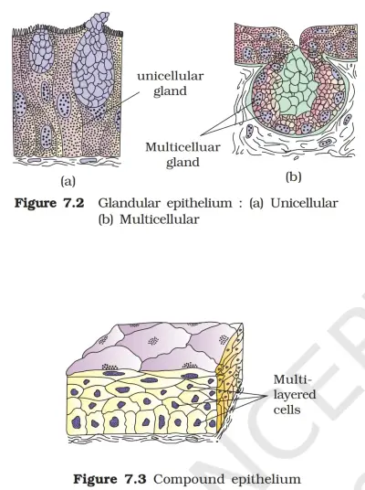
1- Glandular epithelium – Glands are found in this epithelium that produces special chemicals.
What is the gland?
It is a modification of epithelial tissue that produces and secretes certain chemicals.
How many types of glands?
There are many types of glands. There are two types of glands based on cells.
1—Unicellular Glands—A gland in which there is only one cell, and this one cell manufactures and secretes a chemical is called a unicellular gland.
Only two unicellular glands are found in our body.
(i) Goblet cells are found in the innermost layer of our alimentary canal and secrete a thick fluid called mucus. This fluid helps in the forward movement of liquid food by lubricating it.
(ii) Paneth cells – These cells secrete lysozyme in the small intestine, which acts to kill micro-organisms.
2—Multicellular glands—A gland in which many cells together form and secrete a chemical is called a multicellular gland.
Except for the unicellular gland, all other glands are multicellular glands.
Examples – Hypothalamus gland, Pituitary gland, Pancreatic gland, Thyroid gland, Parathyroid gland, Salivary gland, Testes, Ovaries, Adrenal gland, etc.
Gland based on secretion –
Exocrine glands—An exocrine gland transports its secretion or product to its target through its duct.
Examples include the salivary gland, oil gland, sweat gland, lacrimal gland, and milk gland. These glands secrete enzymes and other chemicals.
Endocrine glands do not have a duct and transport their secretion or product to their target through the blood. Examples include the pituitary, hypothalamus, thyroid, thymus, and pineal glands. These glands secrete hormones.
Mixed gland (Heterocrine glands/Mixed glands) – Such a gland that reaches its target through duct and blood is called a mixed gland.
Examples – Pancreas, testis, ovary, etc.
Gland based on shape –
There are three types of glands based on structure.
Tubular glands – These are tubular, hence they are called tubular glands.
Alveolar glands are alveolar in shape; hence, they are called alveolar glands.
Compound Tubulo-Alveolar – In this gland, both tubular and alveolar types of glands are found together; hence, they are called combined tubuloalveolar.
What is a cell junction?
You know what a cell is, and the meaning of junction is joint. Two adjacent cells can join together, which is called a cell junction.
How many types of cell junctions?
This treaty is of three types.
1—Tight Junction—When two cells are firmly connected, this junction type is called a tight junction. It is attached firmly so no substance can leak between the two cells.
2—Adherent junction—This joint also keeps two neighboring cells together but does not stop the leakage of any substance. It has two types.
(i) Interdigitation—Both cell levels are connected to this. For example, if we intertwine our fingers, some are connected like this.
(ii) Desmosomes – In this, there is some protein between the two cells, from which some fibers emerge, which are buried in both the neighboring cells, due to which both the adjacent cells are connected.
3—Gap junction—This junction exchanges essential substances between neighboring cells.
Conclusion
Friends, I hope you have liked the information about epithelial tissue; if you like it, please share it with your friends so they can also benefit. If I have made a mistake in this article, please let me know by commenting.
Thank you